- +91-9244900001
- 40, Pakhowal Rd, New Lajpat Nagar, Model Gram, Ludhiana, Punjab 141001
- 24*7 Emergency
- NABH Accredited
- ECHS Empanelled
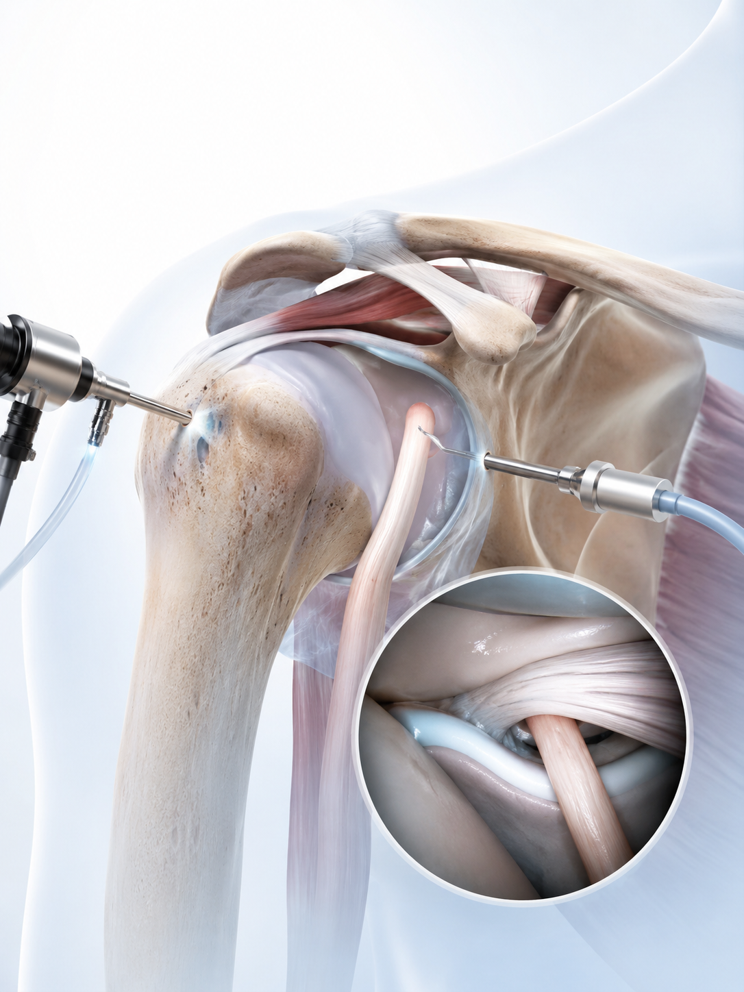
Arthroscopy comes from two words meaning “joint” and “to look” — it is often described to patients simply as keyhole surgery. Rather than making one large incision to open up the shoulder, the surgeon works through a small number of tiny incisions, usually a few millimetres each, known as portals.
An arthroscope — a thin tube fitted with a high-definition camera and a light source — is passed through one portal, projecting a magnified, detailed image of the inside of the joint onto a screen in the operating room. Sterile saline fluid is gently circulated inside the joint throughout the procedure, which gently distends the joint space and keeps the view clear for the surgeon.
Specialised, narrow instruments — probes, shavers, suture passers and anchor devices — are introduced through the remaining portals to examine and treat the joint. Because the surgeon can see the rotator cuff, labrum, cartilage and joint lining directly and in detail, shoulder arthroscopy allows both diagnosis and treatment to happen in the same sitting: if a tear or abnormality is confirmed on inspection, it can often be addressed immediately rather than requiring a separate operation.
Because arthroscopic instruments are narrow and precise, the surrounding muscles, particularly the deltoid, are largely undisturbed. This typically means smaller scars and, in appropriate cases, a faster early rehabilitation compared with many traditional open shoulder procedures — though the overall recovery still depends heavily on which underlying problem was treated.
Many shoulder conditions respond well to a structured non-surgical programme before arthroscopy is ever considered. This may include:
Patient History
Physical Examination
X-rays
MRI
Diagnosis
Discussion of Treatment Options
Individual Treatment Plan
| Condition | Reason for Arthroscopy |
|---|---|
| Rotator Cuff Tear | Repair torn rotator cuff tendons to restore shoulder strength, function, and reduce pain. |
| Bankart Lesion | Repair the torn labrum to restore shoulder stability following recurrent dislocations. |
| Frozen Shoulder | Perform capsular release to improve shoulder mobility and reduce stiffness. |
| Shoulder Impingement | Carry out bursectomy with or without acromioplasty to relieve tendon compression and pain. |
| SLAP Tear | Repair the superior labrum to restore stability and shoulder function. |
| Loose Bodies | Remove loose cartilage or bone fragments causing pain, locking, or restricted movement. |
| Calcific Tendinitis | Remove calcium deposits to relieve pain and improve shoulder movement. |
| Synovitis | Perform synovectomy to remove inflamed synovial tissue and reduce ongoing inflammation. |





Smaller Incisions
Less Soft-Tissue Disruption
Excellent Visualisation
Accurate Diagnosis
Treatment During the Same Procedure
Less Postoperative Pain in Many Patients
Reduced Infection Risk vs Larger Open Incisions
Cosmetically Smaller Scars
Earlier Rehabilitation in Appropriate Cases
| Recovery Stage | Typical Recovery Plan |
|---|---|
| Day of Surgery | Sling for support, regular ice application to reduce swelling, prescribed pain medication, and gentle finger and wrist movements to maintain circulation. |
| Week 1–2 | Focus on wound care, passive shoulder exercises as recommended, and guidance on comfortable sleeping and daily activities. |
| Weeks 2–6 | Begin a supervised physiotherapy programme aimed at gradually improving shoulder range of motion and flexibility. |
| Weeks 6–12 | Progress to a structured strengthening programme targeting the shoulder, rotator cuff, and surrounding muscles. |
| 3–6 Months | Gradual return to sports, manual work, or other demanding activities based on healing, strength, and the specific arthroscopic procedure performed. |
The success of shoulder arthroscopy depends not only on the surgery itself but also on what happens afterward. Patient compliance with the prescribed rehabilitation plan, regular physiotherapy, protecting repaired tendons during the early healing window, performing exercises correctly rather than rushing them, and attending scheduled follow-up visits all directly influence the final outcome. Skipping physiotherapy or returning to activity too early — even after a technically well-performed procedure — can compromise healing, particularly after a rotator cuff or labral repair. At Eva Hospital, a structured, procedure-specific physiotherapy plan is provided alongside surgery, not as an optional add-on.
Dr. Tanveer Bhutani has expertise in the arthroscopic management of sports injuries, rotator cuff disorders and shoulder instability, alongside broader experience in joint preservation and robotic joint replacement. His approach centres on identifying the specific cause of a patient’s shoulder problem through careful clinical assessment, and matching treatment — surgical or non-surgical — to that individual diagnosis rather than applying one standard protocol to every patient.
Shoulder arthroscopy is a minimally invasive keyhole surgery technique that uses a small camera and narrow instruments, inserted through tiny incisions, to examine and treat problems inside the shoulder joint.
Not always. Many knee dislocations cause multi-ligament injury, but multiple ligaments can also be injured without a documented dislocation.
Duration varies with the procedure performed, typically ranging from around 30 minutes for a simple procedure to a few hours for more complex repairs.
It is often performed under general anaesthesia, sometimes combined with a regional nerve block for additional pain control. The anaesthesia team selects the most appropriate approach for each patient.
Many patients undergoing isolated arthroscopic procedures go home the same day, though this depends on the procedure performed and the patient’s overall condition.
Sling duration depends on the procedure — it may be brief after a simple diagnostic arthroscopy or extend to several weeks after a rotator cuff repair.
Driving should resume only once you are no longer impaired by pain medication or a sling and can safely control the vehicle, including emergency manoeuvres.
Consult our specialists for accurate diagnosis and effective treatment solutions.
