
Meniscus Tear Treatment and Surgery in Ludhiana
Meniscus-preserving treatment by Dr. Tanveer Bhutani at Eva Hospital

- What Is the Meniscus?
- Symptoms
- Types of Tears
- Diagnosis
- Can It Heal Without Surgery?
- When Is Surgery Needed?
- Meniscus Repair
- Partial Meniscectomy
- Repair vs Meniscectomy
- Success of Repair
- ACL & Meniscus
- Recovery & Rehab
- Return to Sport
- Risks
- FAQs
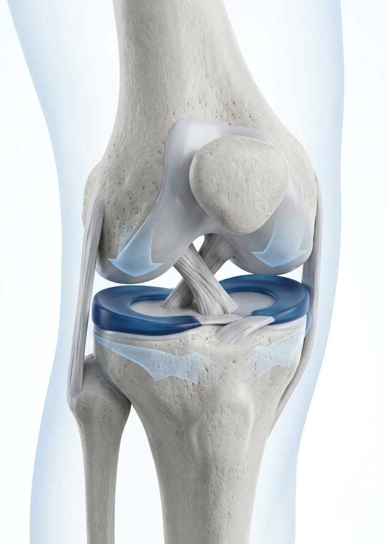
What Is the Meniscus?
The menisci:
- Distribute load across the knee joint
- Absorb shock during walking and impact
- Improve joint stability
- Assist lubrication and nutrition of the joint surfaces
- Protect the articular cartilage
- Contribute to normal knee movement and joint awareness
Meniscus Blood Supply and Healing Zones
Red-Red Zone
Red-White Zone
White-White Zone
How Do Meniscus Tears Occur?
Meniscus tears can occur due to sudden twisting injuries or gradual degeneration over time
Traumatic Meniscus Tears
- Twisting on a planted foot
- Sudden change of direction
- Deep squat or loaded knee flexion
- Football, cricket, basketball, badminton or kabaddi
- Contact injury or ACL injury
- Road traffic trauma
Degenerative Meniscus Tears
- Without one memorable injury
- During routine bending or squatting
- While rising from the floor
- Alongside knee osteoarthritis
- With gradual pain rather than immediate swelling
An MRI finding of a degenerative meniscus tear does not automatically prove that the tear is the main source of pain.
Symptoms of a Meniscus Tear
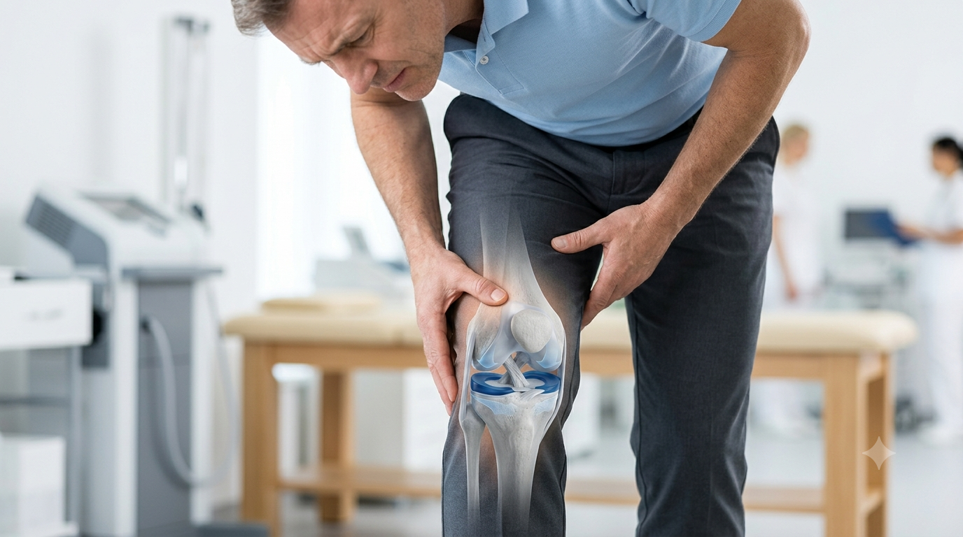
Joint-Line Pain
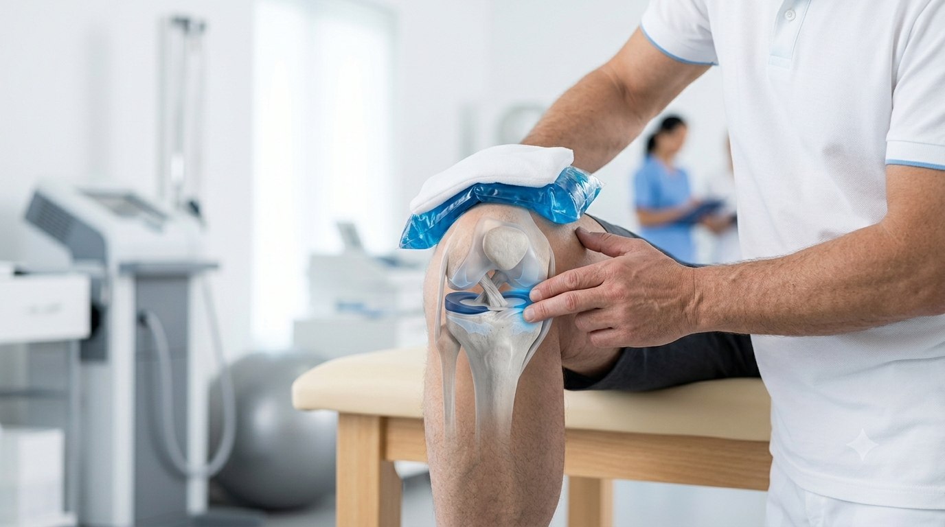
Swelling
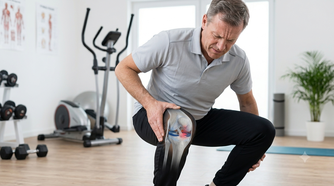
Pain While Squatting
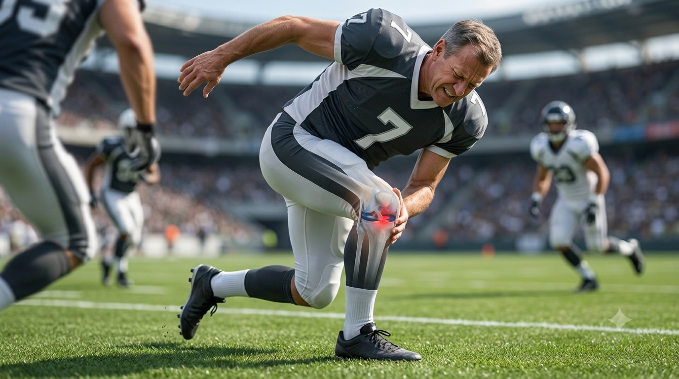
Pain During Twisting
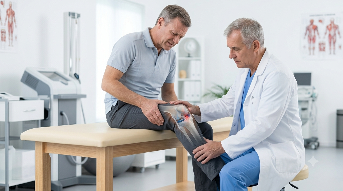
Catching or Clicking
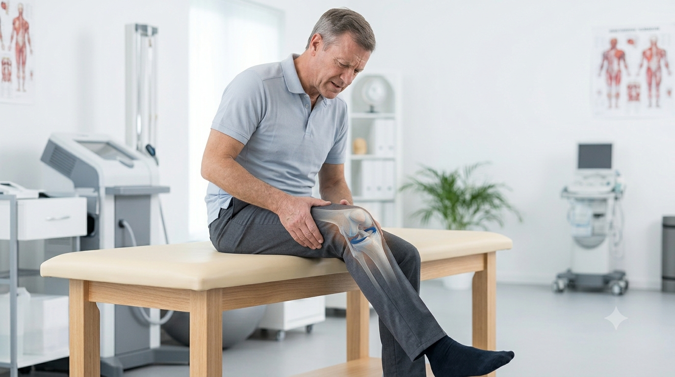
Something Moving Inside
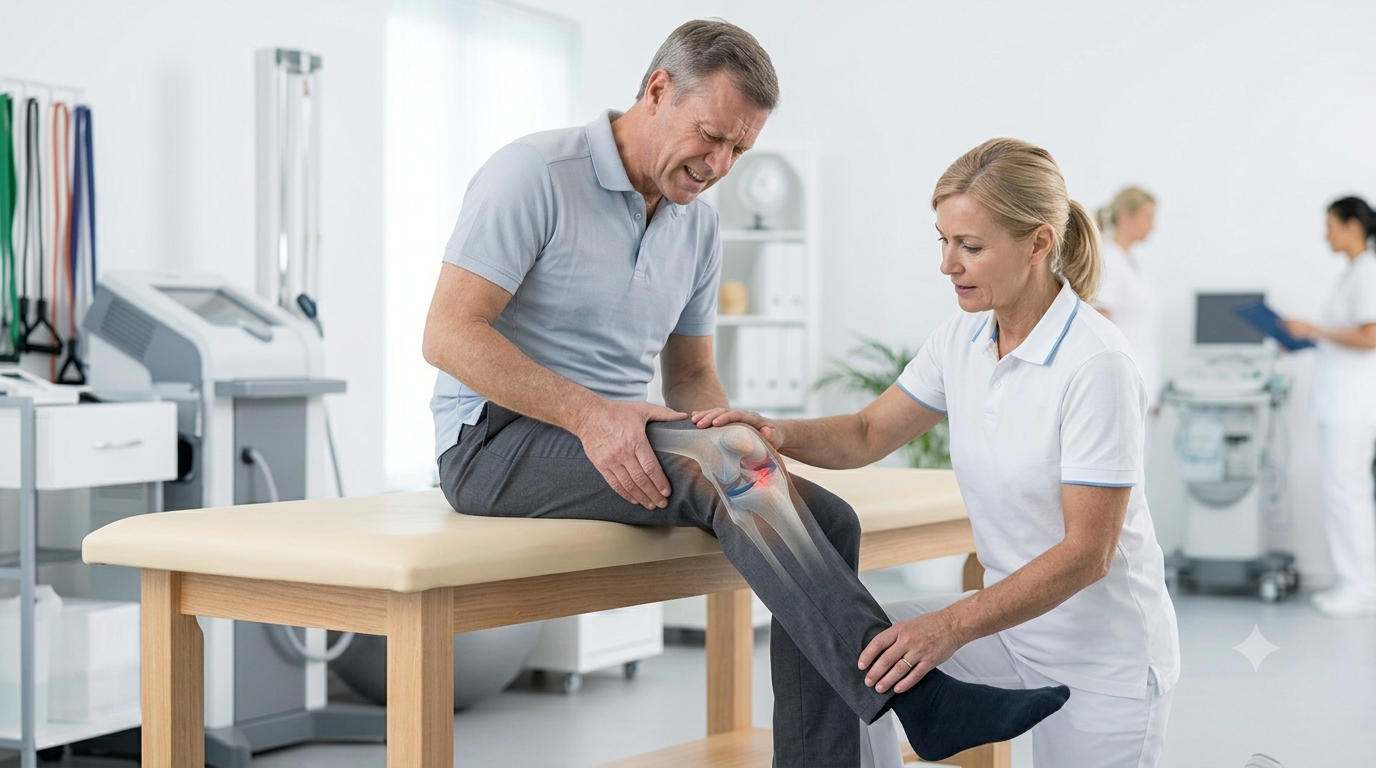
Difficulty Bending/Straightening
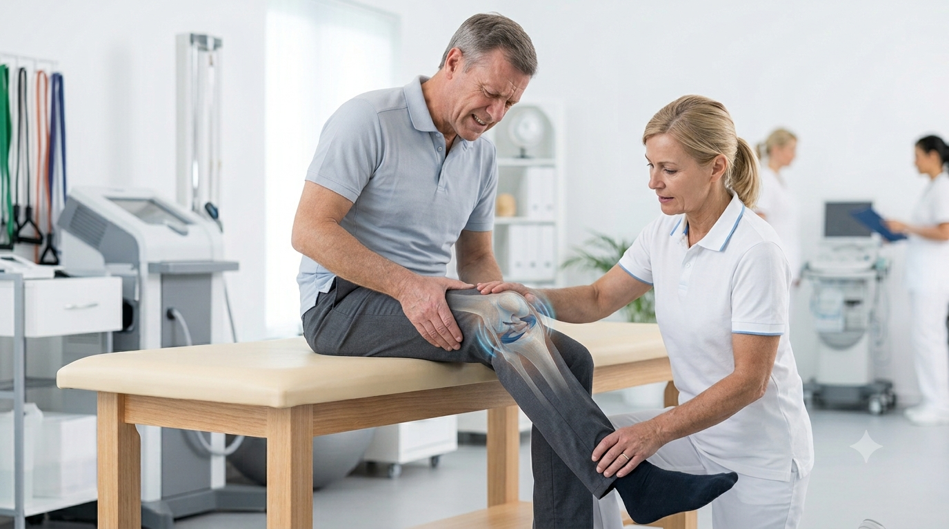
Knee Locking

Giving Way
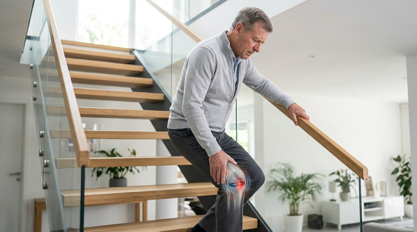
Pain Climbing Stairs
Types of Meniscus Tears
Vertical Longitudinal Tear
Bucket-Handle Tear
Radial Tear
Horizontal Cleavage Tear
Flap or Parrot-Beak Tear
Complex Tear
Meniscal Root Tear
Ramp Lesion
Discoid Meniscus Tear
Medial vs Lateral Meniscus Tears
| Medial Meniscus | Lateral Meniscus |
|---|---|
| Less mobile | More mobile |
| More commonly injured | Frequently injured with acute ACL tears |
| Often associated with twisting injuries and chronic ACL instability | Certain tear patterns can significantly affect rotational stability |
| Posterior-horn tears are common | Preservation is particularly important |
Diagnosis
History of Injury & Symptoms
Clinical Examination
Weight-Bearing X-rays (Where Indicated)
MRI (Suspected Acute Tear or Associated Injury)
Individual Treatment Plan
Clinical Examination
X-rays
MRI
Does Every Meniscus Tear Need Surgery?
Nonsurgical treatment may be suitable when:
Conservative Treatment
- Temporary activity modification
- Ice during the acute phase
- Prescribed pain medication when appropriate
- Physiotherapy and range-of-motion exercises
- Quadriceps, hamstring, hip and core strengthening
- Neuromuscular training
- Gradual return to activity
- Weight management where relevant
Degenerative Tears — A Responsible Note
For many middle-aged or older patients with degenerative, nonlocking meniscus tears, structured exercise-based physiotherapy should be the initial treatment. Long-term trials have found physiotherapy to be noninferior to arthroscopic partial meniscectomy for many degenerative tears.
Anaesthesia and Hospital Stay
What Is Knee Arthroscopy?
Depending on the findings, the surgeon may:
- Repair the tear
- Trim only unstable, irreparable tissue
- Treat an associated ligament or cartilage injury
- Leave stable tissue untouched
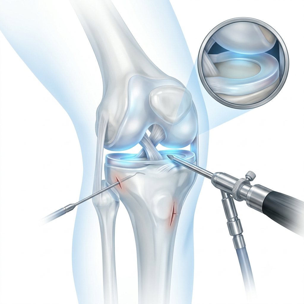
Meniscus Repair
Why Preserve the Meniscus?
- Retaining shock absorption
- Maintaining load distribution
- Protecting articular cartilage
- Supporting knee stability
- Reducing loss of functional meniscal tissue
- Potentially lowering long-term degenerative stress compared with unnecessary tissue removal
When Is Repair More Likely Possible?
- A traumatic tear with good-quality tissue
- A vertical longitudinal tear
- A reducible bucket-handle tear
- A significant radial tear amenable to repair
- A repairable root tear
- A tear in or near a vascular zone
- An associated ACL reconstruction
- A younger or active, compliant patient
Meniscus Repair Techniques
All-Inside Repair
Inside-Out Repair
Outside-In Repair
Meniscal Root Repair
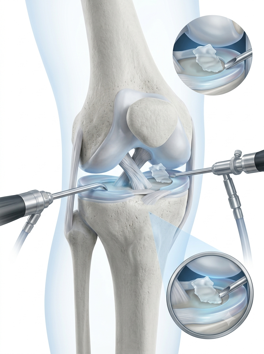
What Is Partial Meniscectomy?
May be appropriate when:
- An unstable symptomatic fragment cannot be repaired
- Degenerated tissue is incapable of holding sutures
- A central avascular tear has poor healing potential
- There is a complex irreparable tear
- Symptoms persist after appropriate nonsurgical treatment
- A flap is producing reproducible mechanical symptoms
Meniscus Repair vs Partial Meniscectomy
| Feature | Meniscus Repair | Partial Meniscectomy |
|---|---|---|
| Aim | Preserve and heal the native meniscus whenever possible. | Remove only irreparable, unstable meniscal tissue while preserving healthy tissue. |
| Suitable Tears | Repairable tears with good healing potential. | Tears that cannot be repaired reliably. |
| Tissue Preserved | Maximum meniscal tissue is preserved. | Some meniscal tissue is permanently removed. |
| Weight Bearing | May require protected weight bearing during early recovery. | Often allows earlier weight bearing, as advised by the surgeon. |
| Movement Restrictions | Frequently required during the initial healing phase. | Usually fewer movement restrictions. |
| Rehabilitation | Longer rehabilitation programme tailored to the tear pattern and repair. | Generally a shorter rehabilitation period. |
| Return to Sport | Commonly takes several months, depending on healing and rehabilitation progress. | Often earlier, depending on symptoms and sporting demands. |
| Risk of Repeat Surgery | Repair may fail or re-tear, occasionally requiring further surgery. | Persistent symptoms or progressive degeneration may require additional treatment. |
| Long-Term Principle | Preferred whenever the meniscus can be repaired reliably to preserve knee function. | Used only when meniscal preservation is not realistically possible. |
What Are the Chances of Meniscus Repair Healing?
Factors Associated With Better Healing
- Peripheral vascular location
- Acute rather than very chronic tear
- Good tissue quality
- Stable reduction and appropriate technique
- Correction of associated ACL instability
- Adherence to rehabilitation
- Avoidance of premature twisting and impact activity
- Nonsmoking status and favourable general health
Factors That May Reduce Healing Potential
- Central avascular location
- Degenerated tissue
- Complex or chronic tear
- Uncorrected instability
- Poor compliance with restrictions
- Reinjury
- Advanced cartilage damage
- Certain root or radial tear characteristics
Biological Augmentation
Meniscus Tears With ACL Injuries
- Meniscus tears frequently accompany ACL injuries
- An unstable ACL can expose the meniscus to repeated injury
- Lateral meniscus tears are common in acute ACL trauma
- Medial meniscus damage may develop with recurrent instability
- Repair can often be performed during ACL reconstruction
- Ramp lesions and root tears must be actively assessed
- Treating the instability may help protect the repaired meniscus
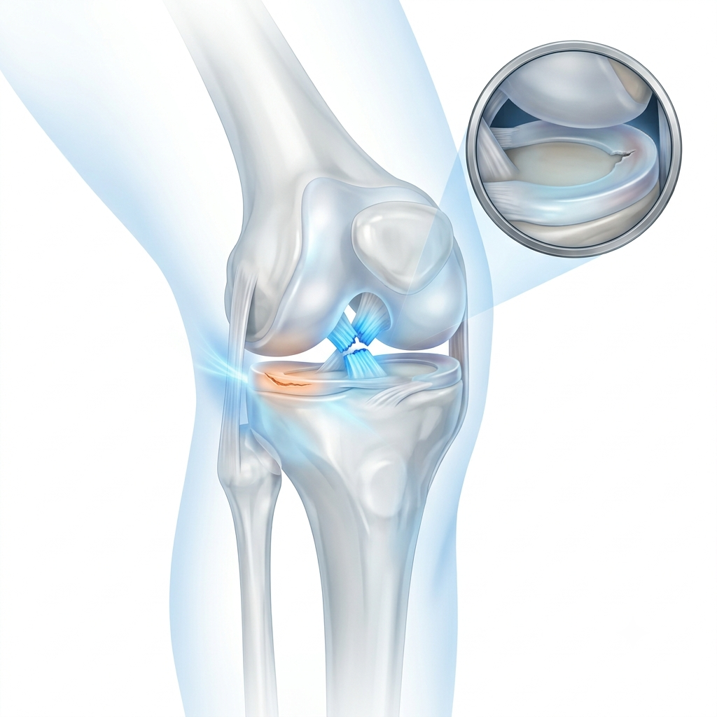
Meniscal Root Tears
Age and activity level
Degree of arthritis
Alignment
Cartilage condition
Body weight
Chronicity of the tear
Ability to follow protected rehabilitation
What Happens on the Day of Surgery?
Admission & Verification
Anaesthesia Assessment
Knee Arthroscopy
Tear & Tissue Assessment
Repair or Selective Meniscectomy
Associated Injury Assessment
Recovery & Physio Instructions
Anaesthesia and Hospital Stay
Anaesthesia is selected after assessment by the anaesthesia team.
Many isolated meniscus procedures are performed as day-care or short-stay surgery.
Admission duration varies with the procedure, associated injuries, medical condition and recovery.
ACL reconstruction with meniscus repair follows a different pathway from isolated meniscus surgery.
Rehabilitation: Meniscectomy vs Repair
After Partial Meniscectomy
- Early phase: swelling control, restoration of movement, quadriceps activation, walking progression as advised.
- Following weeks: strength, balance, stationary cycling, functional exercise, gradual return to work and sport-specific training.
After Meniscus Repair
- Phase 1: protection, brace where prescribed, crutches/protected weight bearing, quadriceps activation, swelling control.
- Phase 2: progressive weight bearing, gradual range of movement, closed-chain strengthening, hip and core conditioning.
- Phase 3: progressive resistance training, balance, single-leg control, cycling.
- Phase 4: jogging only after criteria are met, agility, sport-specific drills, functional testing, gradual return to competition.
Approximate Recovery Comparison
| Activity | Partial Meniscectomy | Meniscus Repair |
|---|---|---|
| Crutches | Often required only briefly, depending on symptoms and comfort. | Commonly needed for a longer period, following the rehabilitation protocol. |
| Weight Bearing | Often allowed early, as advised by the treating surgeon. | May be protected or temporarily restricted during healing. |
| Office Work | Frequently possible within a few days to a few weeks. | Often resumed within a few weeks, depending on mobility and recovery. |
| Driving | When safe knee control, strength, and reaction time have returned. | Usually delayed if bracing or weight-bearing restrictions are still required. |
| Running | Commonly after several weeks once rehabilitation milestones are achieved. | Usually after several months to protect the healing meniscus. |
| Pivoting Sports | Often around 2–3 months in uncomplicated cases with adequate recovery. | Frequently 4–6 months or longer, depending on healing and functional testing. |
| Full Recovery | Commonly achieved within several weeks. | Commonly requires several months of structured rehabilitation. |
Return to Sport
No significant pain or swelling
Full or appropriate range of motion
Strength compared with the opposite side
Single-leg control and balance
Hop or functional tests where appropriate
Confidence and sport-specific movement quality
Surgeon and physiotherapist clearance
Risks and Possible Complications
- Infection
- Blood clot
- Stiffness
- Persistent pain
- Swelling
- Nerve or blood-vessel injury
- Failure of repair to heal
- Re-tear
- Need for repeat arthroscopy
- Further meniscal tissue loss
- Persistent symptoms from cartilage damage or arthritis
- Anaesthetic complications
Why Can Pain Continue After Meniscus Surgery?
Coexisting osteoarthritis
Cartilage injury
Bone marrow oedema
Muscle weakness
Incomplete rehabilitation
Persistent swelling
Re-tear or failed repair
The MRI tear was not the sole pain generator
Other knee pathology
Why Choose Dr. Tanveer Bhutani?
Preservation Whenever Possible
Nonsurgical First, When Appropriate
Individualised Technique
Procedure-Specific Rehab
Patient Journey at Eva Hospital
Consultation
Clinical Examination
X-rays / MRI When Indicated
Conservative Care or Surgery Discussion
Meniscus-Preserving Plan
Procedure When Required
Physiotherapy
Return-to-Work/Sport Assessment
Patient Stories
| Age Group | Typical Meniscus Injury & Management |
|---|---|
| 19–24 Years |
Young Athlete – Repairable Bucket-Handle Tear Symptoms: Knee locking after a twisting football injury. Treatment: Arthroscopic reduction with meniscus repair to preserve cartilage. Outcome: Successfully returned to competitive sports after structured rehabilitation. |
| 20–28 Years |
ACL Reconstruction with Meniscus Repair Symptoms: Knee instability associated with a lateral meniscus tear. Treatment: Combined ACL reconstruction and arthroscopic meniscus repair. Outcome: Returned to sport after completing strength and functional testing. |
| 30–40 Years |
Meniscal Root Tear Symptoms: Deep knee pain with early degenerative cartilage changes. Treatment: Arthroscopic meniscal root repair followed by protected rehabilitation. Outcome: Improved knee function with continued long-term follow-up. |
| 45–55 Years |
Irreparable Flap Tear Symptoms: Mechanical catching caused by an unstable meniscal flap. Treatment: Selective arthroscopic partial meniscectomy while preserving healthy meniscus tissue. Outcome: Relief of mechanical symptoms with return to normal daily activities. |
| 55–65 Years |
Degenerative Meniscus Tear Symptoms: Gradual onset of knee pain without true locking episodes. Treatment: Structured physiotherapy, strengthening exercises, weight management, and activity modification. Outcome: Significant symptom improvement without requiring surgery. |
Related Pages
Frequently Asked Questions
Some tears may settle or heal, particularly stable peripheral tears with a better blood supply. Other tears may remain visible on MRI but become asymptomatic. Treatment depends on symptoms, tear type, stability and patient factors.
No. Meniscus tears, particularly degenerative tears, can appear on MRI without being the main source of pain.
Physiotherapy does not stitch torn tissue together, but it can reduce pain, restore strength and function, and allow many patients to avoid surgery.
Prompt assessment is advisable when a displaced tear causes true locking, substantial loss of movement or a suspected repairable bucket-handle tear.
Repair preserves the meniscus by suturing a suitable tear. Partial meniscectomy removes only unstable tissue that cannot be repaired reliably.
Repair is preferred when the tear has realistic healing potential. Attempting to repair severely degenerated or nonviable tissue may expose the patient to a prolonged recovery and later failure.
The decision is based on tear pattern, location, blood supply, tissue quality, chronicity, stability, associated injuries and patient factors. Final tissue assessment may occur during arthroscopy.
Many modern repairs remain successful in roughly 75–85% of appropriately selected patients, but success varies and cannot be guaranteed.
Selected recurrent tears may be suitable for revision repair. Others may require limited partial meniscectomy.
Postoperative discomfort and swelling are expected, but pain is managed through medication, ice, activity guidance and rehabilitation.
Not every patient needs one. A brace may be prescribed after specific repairs or associated ligament surgery.
Crutches may be needed briefly after partial meniscectomy and generally for longer after certain repairs. Instructions depend on the procedure.
Walking instructions depend on whether the meniscus was repaired or trimmed and on the tear pattern. Some repairs require protected weight bearing.
Desk work may resume earlier than work involving prolonged standing, climbing, squatting or lifting.
Driving is appropriate only when the patient is no longer impaired by medication or a brace and can control the vehicle safely, including emergency braking.
Upper-body and selected controlled exercises may resume earlier. Running, deep squats and twisting require clearance according to the procedure.
Squatting is usually reintroduced gradually. Deep loaded flexion may be restricted during early healing.
Many patients return to sport after successful treatment and rehabilitation. Clearance should be based on healing, strength, control and sport-specific testing.
It is a displaced longitudinal tear that may block knee movement and cause locking.
It is a tear near the meniscus attachment to the tibia that can severely impair meniscal function.
Yes. Meniscal injuries commonly accompany ACL tears and may be repaired during ACL reconstruction.
Loss of meniscal tissue increases joint-surface loading and is associated with degenerative risk. The amount, location and condition of the knee all matter, which is why tissue preservation is prioritised.
PRP may be considered as an adjunct in selected situations, but evidence is variable and it does not reliably heal every tear or replace surgery when a displaced repairable tear requires fixation.
It uses small incisions, but it remains a surgical procedure with anaesthetic, rehabilitation and complication considerations.
A repaired meniscus can fail or re-tear, and remaining meniscal tissue can sustain a new injury after either repair or meniscectomy.
Some tears remain stable or become asymptomatic. Others may continue causing pain, locking or secondary damage. The risk depends on the tear pattern, symptoms and stability.
Usually not as the first treatment when there is no true locking. Exercise-based rehabilitation is commonly preferred initially.
Not necessarily. Pain due to osteoarthritis, cartilage damage or other pathology may persist even if the meniscus tear is treated correctly.