MPFL Reconstruction for Recurrent Patellar Dislocation in Ludhiana

Individualised treatment of kneecap instability by Dr. Tanveer Bhutani at Eva Hospital, Ludhiana
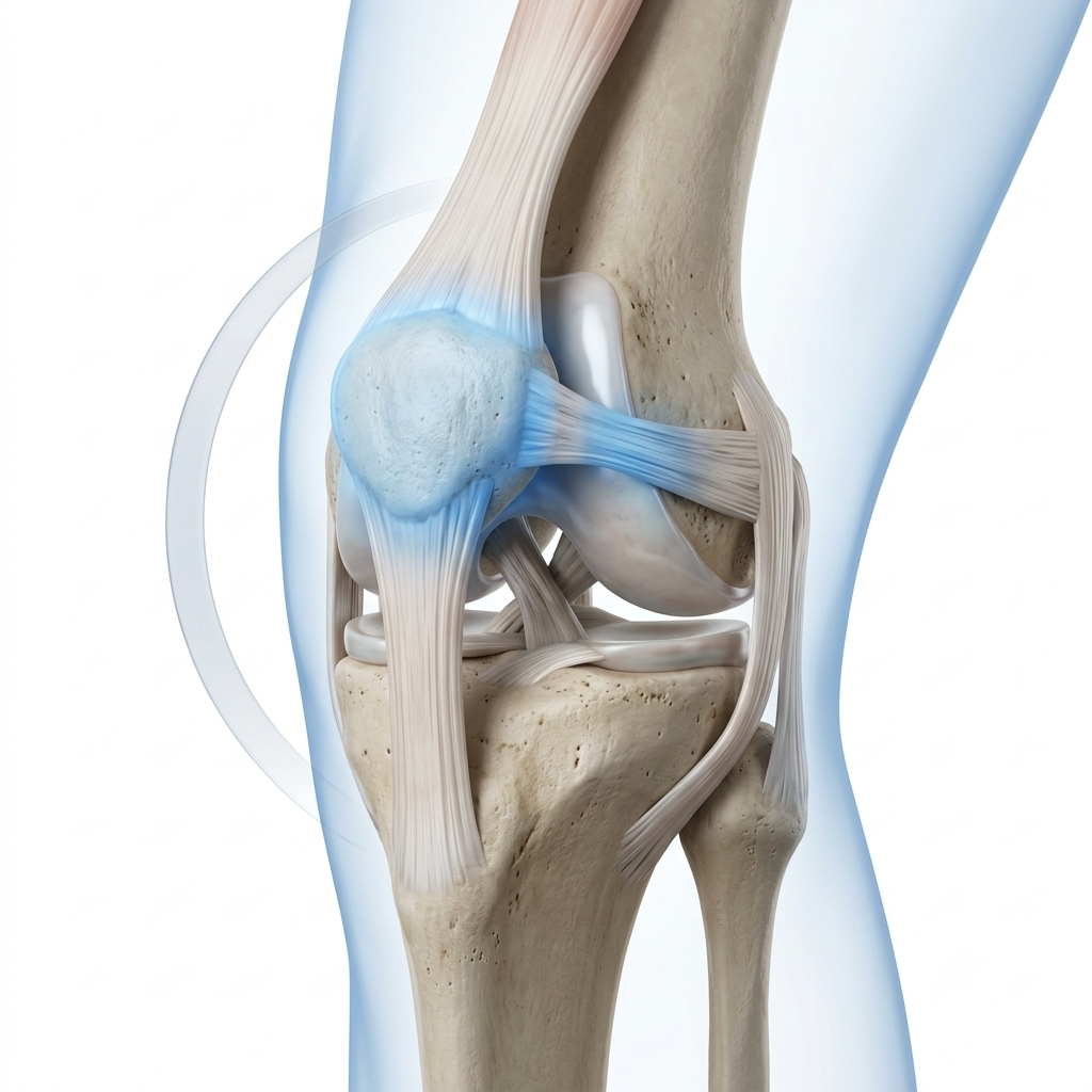
The kneecap may dislocate during a twisting injury, sports activity, fall or direct impact. While many first-time patellar dislocations can be treated without surgery, repeated dislocations or persistent instability can make the knee unreliable and may damage the cartilage beneath the kneecap.The medial patellofemoral ligament — or MPFL — is an important restraint that helps prevent the kneecap from moving excessively toward the outside of the knee. MPFL reconstruction may be considered for selected patients with recurrent patellar instability, after detailed assessment of the ligament, cartilage, knee alignment, trochlear shape and other anatomical risk factors.At Eva Hospital, Dr. Tanveer Bhutani individualises treatment rather than recommending one standard operation for every dislocating kneecap.
Dr Tanveer bhutani Ortho surgeon

Understanding the Kneecap

The patella (kneecap) normally moves within the trochlear groove at the lower end of the femur. The quadriceps muscle and tendon, patellar tendon, trochlear groove, limb alignment and medial soft tissues collectively guide and stabilise it.

What Is the MPFL?

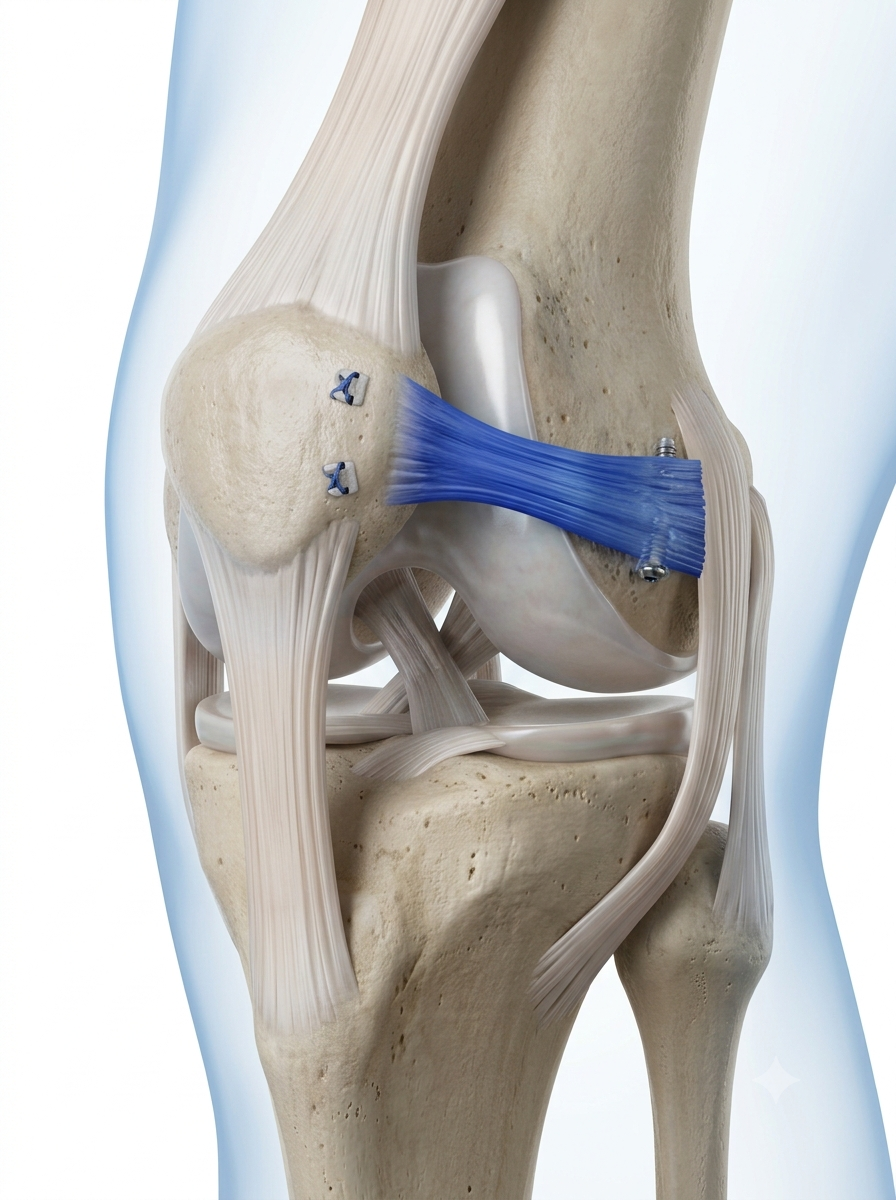
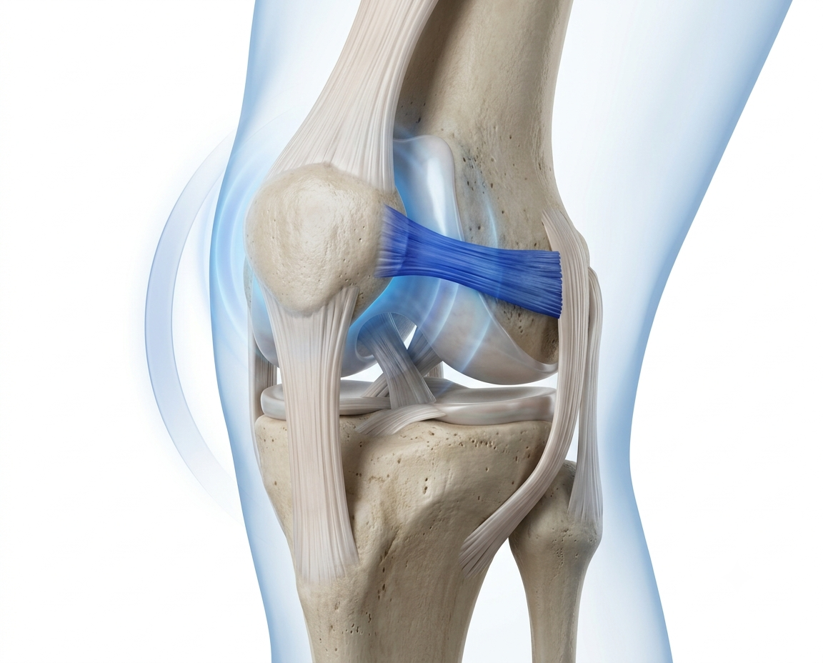
The medial patellofemoral ligament connects the inner aspect of the kneecap to the inner side of the femur. It is an important restraint against outward displacement of the patella, especially during the early part of knee bending.

The MPFL is commonly stretched or torn during a lateral patellar dislocation. In some patients, it heals sufficiently with rehabilitation. In others — particularly those with recurrent instability — it may no longer provide adequate restraint.
How Does a Patellar Dislocation Occur?

Injury Mechanisms

Anatomical Predisposition

Symptoms

Sudden Kneecap Displacement
Visible Deformity (Acute)
Pain Around the Kneecap
Rapid Swelling
Inner Kneecap Tenderness
Feeling the Patella May Move Out
Repeated Subluxation
Recurrent Dislocation
Apprehension During Bending
Difficulty With Stairs
Difficulty Running / Changing Direction
Clicking or Catching

Circulation and nerve examination

X-rays

Swelling and tenderness assessment

Evaluation for fracture

MRI where indicated

Temporary brace or support

Crutches

Orthopaedic follow-up

First-Time vs Recurrent Dislocation

First-Time Patellar Dislocation

Many first-time dislocations can be treated without immediate reconstruction, provided:

Recurrent Dislocation

Repeated dislocation or persistent subluxation may indicate that the medial stabilising structures no longer provide sufficient control. It can lead to:

Diagnosis

History of Dislocation / Instability

Clinical Examination

X-rays

MRI

Alignment / Rotational Imaging (When Indicated)

Individual Treatment Plan

Clinical Examination

Patellar apprehension, direction and degree of patellar movement, tracking, J-sign, knee alignment, generalised laxity, quadriceps and hip control, range of movement, ACL and other ligament stability, meniscus/cartilage symptoms.

X-rays

Patellar position, trochlear shape, patellar tilt, patella alta, fracture, osteochondral fragment, joint alignment.

MRI

MPFL injury pattern, cartilage damage, bone bruising, osteochondral fracture, loose body, trochlear anatomy, other ligament or meniscus injury.

CT / Alignment Imaging

May be considered when assessment of rotational alignment, tibial-tubercle position or bony anatomy is required.

Can Patellar Instability Be Treated Without Surgery?

Physiotherapy May Include

Bracing and Taping

A brace or taping may provide short-term support in selected cases but should not replace muscle restoration and movement retraining.

When May Surgery Be Recommended?

MPFL reconstruction may be considered when there is:
Recurrent lateral patellar dislocation
Repeated symptomatic subluxation
Persistent objective instability
Failure of appropriate rehabilitation
Significant apprehension restricting normal activity
Instability affecting sport, work or daily life
A suitable anatomical pattern for isolated reconstruction
Associated pathology requiring surgical treatment

When Is Isolated MPFL Reconstruction Sufficient?

Recurrent instability is the primary complaint

The MPFL is functionally insufficient

Bony alignment does not require separate correction

Trochlear or patellar-height abnormalities are not judged to require another operation

Cartilage damage does not require a different treatment strategy

Skeletal maturity and anatomy permit the planned reconstruction

Associated Procedures

Osteochondral-Fragment Treatment

A loose or displaced cartilage-and-bone fragment may need fixation or removal, depending on size, location and viability.

Tibial Tubercle Osteotomy

The tibial tubercle may be repositioned in selected patients to address abnormal tracking, alignment, patellar height or joint loading.

Trochleoplasty

Reshaping of the trochlear groove is reserved for selected severe cases — not routinely required for every patient with trochlear dysplasia.

Cartilage Treatment

Cartilage injury may be treated during the same surgery when clinically appropriate.

Lateral-Sided Procedures

Used only for selected indications — not performed automatically for instability.

Skeletally Immature Patients

Surgical planning must protect the growth plates; techniques may differ in children and adolescents who have not completed growth.

What Is MPFL Reconstruction?

MPFL reconstruction recreates the function of the damaged medial patellofemoral ligament using a tendon graft. The graft is attached to the kneecap and femur at anatomically planned locations, with the aim of providing stability while allowing normal patellar movement.

Goals:

Graft Options

Gracilis Tendon
Semitendinosus Tendon
Quadriceps Tendon
Other Autograft Options
Allograft (Selected Situations)

Fixation Techniques

Fixation may involve suture anchors, patellar sockets or tunnels, soft-tissue fixation, femoral socket or tunnel fixation, and implant-free or limited-implant techniques in selected cases.

What Happens During Surgery?

Anaesthesia & Examination

Arthroscopic Assessment (Where Indicated)

Loose Body / Cartilage Treatment

Graft Preparation

Anatomical Fixation

Tracking & Tension Check

Closure & Rehab Plan

Not every patient requires arthroscopy or an associated bony procedure.

Hospital Stay and Pain Management

Anaesthesia evaluation
Multimodal pain management
Swelling control
Early physiotherapy
Brace where prescribed
Crutch training
Discharge after safe mobilisation

Rehabilitation After MPFL Reconstruction

Protection & Swelling Control

Protect the reconstruction, control pain and swelling, restore quadriceps activation, begin safe knee movement, brace/crutches as advised, avoid falls and twisting.

Restore Movement & Walking

Progressive range of movement, gradual weight-bearing, patellar mobility where appropriate, normal walking pattern, controlled closed-chain strengthening.

Strength & Control

Quadriceps, hip and core strengthening, balance, single-leg control, stationary cycling, progressive functional exercises.

Running & Agility

Running only after strength, control and swelling criteria are met; progressive jumping/landing; directional-change drills; sport-specific movement; confidence assessment.

Return to Sport

Functional testing, strength comparison, movement-quality assessment, no significant pain/swelling, no recurrent apprehension, surgeon and physio clearance.

Individualised Adjustments

Protocols differ if MPFL reconstruction is combined with tibial tubercle osteotomy, cartilage fixation, osteochondral surgery, trochleoplasty, or another ligament procedure.

Approximate Recovery Milestones

MilestoneApproximate Expectation
Crutches and BraceDuration depends on the surgical procedure performed and the rehabilitation protocol prescribed by the surgeon.
Desk-Based WorkMany patients are able to return within a few weeks, depending on comfort, mobility, and job demands.
Comfortable Routine WalkingUsually achieved progressively during the early stages of rehabilitation as strength and confidence improve.
DrivingCan resume once safe vehicle control, reaction time, and knee function have been restored.
CyclingTypically introduced during rehabilitation when adequate knee movement and control have been achieved.
JoggingCommonly begins after several months, following satisfactory clinical assessment and functional clearance.
Sports TrainingOften resumes around 6–9 months, depending on recovery progress and successful functional testing.
Full Pivoting SportFrequently permitted after 9 months or longer, once strength, stability, and sport-specific criteria are met.

Expected Results

Most appropriately selected patients experience improved stability and fewer recurrent dislocation episodes after MPFL reconstruction. However, outcomes depend on correct identification of all contributing anatomical factors, accurate graft positioning, appropriate tensioning, associated cartilage damage and rehabilitation. Published reviews generally report low recurrence after isolated reconstruction, but results vary between studies and patient groups. A modern systematic review found recurrent instability ranging from 0% to approximately 11%, while complications varied considerably according to technique and study definition.

Risks and Complications

Why Might Surgery Fail?

Unrecognised bony risk factors
Incorrect diagnosis
Graft malposition
Excessive or insufficient graft tension
Significant patella alta
Severe trochlear dysplasia
Rotational malalignment
Reinjury
Inadequate rehabilitation
Generalised ligamentous laxity
Advanced cartilage damage
Premature return to sport
Successful treatment requires more than simply reconstructing one ligament.

Why Choose Dr. Tanveer Bhutani?

Recurrent patellar instability requires assessment of the entire patellofemoral joint — not only the torn MPFL. Dr. Tanveer Bhutani evaluates the patient’s dislocation history, patellar tracking, trochlear shape, patellar height, alignment, cartilage condition, skeletal maturity and sporting requirements before recommending treatment.

Clinical & Imaging Evaluation

A full patellofemoral assessment, not just an MRI report.

Nonsurgical Treatment First

Surgery is not the default for every dislocation.

Individualised Reconstruction

Assessment for associated bony or cartilage procedures when needed.

Structured Follow-Up

Physiotherapy, return-to-sport planning and long-term review.

Patient Journey

Consultation

Patellar Stability & Alignment Exam

X-rays & MRI

Physio or Surgical Discussion

Individualised Procedure

Protected Rehabilitation

Strength & Functional Testing

Return to Activity

About Your Surgeon

Dr. Tanveer Bhutani

Dr. Tanveer Bhutani treats patellofemoral instability, ligament injuries and complex sports trauma at Eva Hospital, Ludhiana, with an approach centred on individualised, evidence-based assessment rather than one standard operation for every patient.

Related Pages

Sports Injuries

Our full sports medicine and ligament reconstruction service.

Knee Arthroscopy

How minimally invasive knee surgery works.

ACL Reconstruction

Our complete guide to the anterior cruciate ligament.

PCL Reconstruction

Understanding posterior cruciate ligament injuries.

Meniscus Surgery

Preservation-first meniscus treatment.

Multi-Ligament Reconstruction

Managing complex, multi-structure knee trauma.

Physiotherapy & Rehabilitation

Structured, procedure-specific recovery programmes.

Fracture & Trauma Care

Management of associated fractures and acute trauma.

Frequently Asked Questions

The MPFL is an important ligament on the inner side of the kneecap that helps prevent excessive outward movement.
It is commonly stretched or torn, but the exact injury location and severity vary.
No. Many first-time dislocations are initially treated with rehabilitation unless there is an associated fracture, loose body or another compelling indication.
Its principal indication is recurrent symptomatic lateral patellar instability.
Physiotherapy can improve strength and movement control and is effective for many patients, but it cannot correct every anatomical or structural cause of instability.
Subluxation is partial displacement; dislocation means the kneecap completely leaves the trochlear groove.
It is an abnormal or shallow shape of the groove in which the kneecap normally travels.
It means the kneecap sits higher than usual, which can delay its engagement within the trochlear groove during bending.
It is a procedure that repositions the attachment of the patellar tendon in selected patients to improve alignment, tracking or loading.
No. Additional bony procedures are used only when the patient’s anatomy and instability pattern justify them.
Common options include gracilis, semitendinosus or quadriceps tendon. Selection is individualised.
It may be used to evaluate or treat cartilage damage, loose bodies or other internal knee pathology when indicated.
Many patients receive a brace initially, but the duration depends on the procedure and rehabilitation plan.
Walking usually begins with protection and assistive support, progressing according to the reconstruction and any associated procedures.
Driving should resume only when the patient is no longer impaired by medication or a brace and can safely control the vehicle.
Many patients return within a few weeks, depending on pain, mobility, transport and the nature of their work.
Running is introduced only after adequate movement, strength, stability and functional control have returned.
Many patients return to sport after successful reconstruction and rehabilitation, but clearance is based on objective recovery rather than time alone.
Yes, although recurrent instability is relatively uncommon in appropriately selected and treated patients.
Not necessarily. The operation primarily treats instability. Pain from cartilage damage, overload or other patellofemoral conditions may persist.
Yes, but the technique must account for open growth plates and skeletal development.
Repeated episodes can cause fear, functional restriction and further cartilage or osteochondral injury.

Dealing With a Recurrently Unstable Kneecap?

Book an orthopaedic evaluation with Dr. Tanveer Bhutani at Eva Hospital, Ludhiana.
Hip Replacement

Send Message

Consult our specialists for accurate diagnosis and effective treatment solutions.

pop up