 Eva Hospital
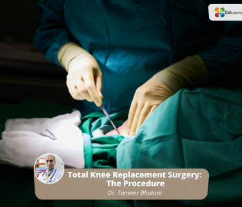
Total Knee Replacement Surgery: The Procedure
What is Knee Replacement Surgery?
Total Knee replacement surgery or knee arthroplasty is a surgical procedure that helps to relieve pain and restore function in knee joints, that are damaged by arthritis, and cannot be controlled by other treatments.
The total knee replacement procedure includes cutting away damaged bone and cartilage from the thigh bone, shinbone and kneecap to replace it with an artificial joint called a prosthesis which is made of metal alloys, high-grade plastics, and polymers.
A surgeon can best assess the knee’s range of motion, stability, and strength through physical examination and with help of X-rays to determine whether Knee Replacement is a viable option for you.
Reasons for the Knee Replacement Procedure
Various types of arthritis can affect the knee joint:
- Osteoarthritis
- Rheumatoid arthritis
- Traumatic arthritis
Osteoarthritis causes the breakdown of joint cartilage, leading to damage of the cartilage and bones limiting the movement and causing pain.
Acute degenerative joint disease can cause pain and swelling, making it difficult for people to do normal activities that include bending the knee like walking or climbing stairs.
Knee replacement surgery is generally done as a treatment to relieve pain and disability in the knee. The most common cause of knee replacement surgery is osteoarthritis.
In some cases, a knee injury can cause rheumatoid arthritis and arthritis leading to a degeneration of the knee joint. Moreover, fractures, torn cartilage, and torn ligaments may sometimes lead to irreversible damage to the knee joint.
Knee replacement surgery can prove to be an effective treatment when other treatment options like anti-inflammatory medication, physical therapy, and cortisone injections into the knee joint fail to alleviate the pain.
Anatomy of the Knee Replacement
Before we try to understand the Knee replacement procedure, it is important to understand the anatomy of the knee.
The knee consists of:
- Tibia, the shin bone or larger bone of the lower leg.
- The femur, the thigh bone, or the upper leg bone.
- Patella, also known as the kneecap.
- Cartilage, a type of tissue covering the surface of a bone at a joint, helps to absorb shock and protect the knee and reduce movement within a joint.
- The synovial membrane is a tissue lining the joint and sealing it into a joint capsule. It also secretes synovial fluid which is a clear, sticky fluid found around the joint to lubricate it.
- Ligaments are a type of strong, elastic bands of connective tissue that surround the joint to give assistance to the knee and to help limit the joint’s movement. It connects bone to the bone.
- Tendon, the tough cords of connective tissue that connect muscles to bones and help to control the movement of the joint.
- The meniscus is a curved part of cartilage in the knees and other joints that act as a shock absorber, increasing contact area. They help to deepen the knee joint.
Knee Replacement procedure
Before the Procedure:
- A complete physical examination is done to ensure the patient is in good health before undergoing the procedure. Blood tests or other diagnostic tests are also performed.
- Patients should inform the doctor if they are sensitive or allergic to any medications, latex, tape, and anesthetic agents.
- All regular medications being taken by the patient and medical conditions if any should also be conveyed to the doctor.
- The patient is asked to fast for eight hours before the procedure.
- A sedative is given before the procedure to help the patient relax.
- One should also make arrangements before the surgery itself, for someone to help around the house for a week or two after being discharged from the hospital.
- The decision about the type of anesthesia to be used is also made after a discussion between the anesthesiologist and the patient.
- Intravenous antibiotics are given before, during, and after the procedure to help prevent post-surgical infections.
During the Procedure knee Replacement
Knee replacement surgery is usually performed under general anesthesia. However, all options are discussed with the patient in advance by the anesthesiologist.
The anesthesiologist continuously monitors the heart rate, blood pressure, breathing, and blood oxygen level during the surgery.
The actual procedure of the surgery is as follow:
Step 1: Making the knee incision
An incision of about 8 to 10 inches long is made by the surgeon across the front of the knee to gain access to the patella or kneecap.
Step 2: Rotating the patella (kneecap)
Once the knee is open, the patella is rotated outside the knee area, allowing the surgeon to view the area needed to perform the surgical procedure.
Step 3: Preparing the femur (thighbone)
The femur known as the thighbone is the first bone to be surfaced. Bones are carefully measured by the surgeon and precise cuts are made using special instruments.
The damaged joint surfaces are cut away. The surgeon then cuts and resurfaces the end of the femur to fit the first part of the artificial knee, the femoral component.
The prosthesis is normally composed of 3 components: the tibial component, the femoral component, and the patellar component
Step 4: Implanting the femoral component
The metal femoral component or artificial joint is then attached to the end of the femur and bone cement is used to seal it into place.
Step 5: Preparing the tibia (shinbone)
The next bone to be resurfaced is the tibia or shinbone. The damaged bone and cartilage are removed from the top of the tibia and then the bone is shaped to fit the metal and plastic tibial components.
Step 6: Implanting the tibial component
The tibial tray, which is the bottom portion of the implant, is fitted to the tibia and secured into place using bone cement.
Once it is in place, the surgeon snaps in a polyethylene insert made of medical-grade plastic to sit between the tibial tray and the femoral component, which acts as a kind of buffer.
This insert will eventually provide support for the body as one bends and flexes the knee.
Step 7: Re-adjusting the patella
The surgeon sometimes needs to flatten the patella before returning it to its normal position and fit it with an additional plastic component to ensure a proper fit with the rest of the implant. The plastic piece is sometimes cemented to the underlying bone if the surgeon feels the need.
Step 8: Finalizing the procedure
The knee is bent and flexed to ensure that the implant is working correctly and to check if the alignment, sizing, and positioning are suitable.
The procedure is completed by closing the incision with stitches or staples, and then bandaging it, and preparing the patient for recovery.
One might leave the operating room with the leg in a CPM (continuous passive motion) machine that gently bends and flexes the new knee while the patient is lying down.
The Total Surgery lasts about two hours.
After the procedure Knee Replacement
The patient is taken to the recovery room for observation after the surgery. The patient is shifted to the hospital room once the blood pressure, pulse, and breathing are stable and the patient is alert. There is a hospital stay of a few days after the surgery.
Starting to move the new joint after surgery is very important. A physical therapist plans an exercise program after the surgery.
The pain is controlled with adequate medication so that the patient is comfortable and can exercise. An exercise plan to follow both in the hospital and after discharge is provided by the physical therapist until one regains muscle strength and a good range of motion.
Home Care
All kinds of falls must be avoided after the knee replacement surgery, as it can result in damage to the new joint. A cane or walker is recommended until the strength and balance improve.
Certain modifications around the home can help one during the recovery. Some of these include:
- Handrails along all stairs
- Safety bars or handrails in the shower or bath
- Using a bench or chair in the shower
- Toilet seat riser with arms
- Reaching stick to pick objects
- Removing all kinds of loose carpets and electrical cords that may cause one to trip or fall
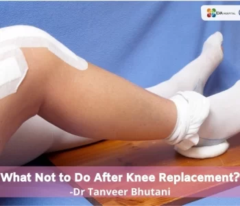
Also Read:- What Not To Do After Knee Replacement?
Doctor’s Note
Dr. Tanveer Bhutani at Eva Hospital who specializes in the best Joint Replacement surgeon in Ludhiana says that total knee replacement surgery provides pain relief and helps in improving mobility, providing a better quality of life.
He adds that when done by a competent surgeon and through a correct procedure, most knee replacements can be expected to last for more than 15 years.
One can usually resume most of the daily activities, 3 to 6 weeks after the surgery and Driving is also feasible at around three weeks if one can bend the knee far enough to sit in a car, and has sufficient muscle control to operate the brakes and accelerator.
One can engage in many low-impact activities, like walking, swimming, or biking, after complete recovery. However, higher impact activities such as jogging, skiing, tennis, and sports that involve contact or jumping should be avoided.
So if you have let the knee pain hinder your normal activities for too long, it is time to consider Total Knee Replacement surgery which can help give you a painful life, allowing you to engage in activities that give you joy, pleasure, and happiness.
Dr. Tanveer Bhutani Orthopedic Specialist has have been helping people lead pain-free and happy lives for several years now. Contact to book an appointment to get your condition accessed and about a discussion of your treatment options.
